


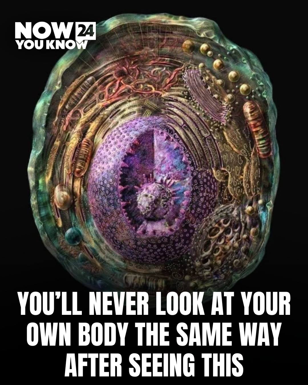
A breathtaking scientific visualization is going viral online after revealing what it actually looks like inside a living human cell using real molecular data.
The reconstruction combines advanced imaging techniques with cinematic 3D rendering, creating an experience many viewers described as “looking into another universe.”
Built Using Real Scientific Data
Unlike artistic interpretations often seen in textbooks or animations, this reconstruction is based on actual molecular and structural data gathered through X-ray crystallography and electron microscopy.

These technologies allow scientists to map proteins, cellular structures, and microscopic interactions at incredibly detailed levels.
Researchers then use that information to digitally rebuild the environment inside a cell as accurately as possible.
Why the Visualization Feels So Real
Part of what makes the reconstruction so striking is the use of light, transparency, and movement.
Instead of presenting cells as flat diagrams, the visualization creates a layered 3D environment that gives viewers the sensation of physically moving through the inside of a living cell.
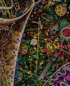
Scientists say this approach helps people better understand just how crowded, dynamic, and active microscopic life actually is.
The Inside of a Cell Is Constantly Moving
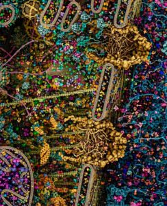
Experts explain that cells are far from static structures.
Inside every human cell, proteins, molecules, and microscopic machines are constantly transporting information, generating energy, repairing damage, and communicating with one another at astonishing speed.
The visualization captures that nonstop activity in a way traditional scientific illustrations often struggle to show.
X-Ray Crystallography and Electron Microscopy Explained
X-ray crystallography works by analyzing how X-rays scatter when they pass through crystallized molecules, allowing scientists to determine detailed molecular structures.
Electron microscopy, meanwhile, uses beams of electrons instead of light to capture images at resolutions powerful enough to reveal structures far too small for ordinary microscopes.
Together, the technologies have transformed modern biology and medicine by giving researchers unprecedented views inside living systems.
Why Scientists Believe Visualizations Like This Matter
Researchers say turning scientific data into immersive visual experiences can help both students and the public better understand complex biological processes.
Many people online admitted they finally grasped how alive and intricate cells truly are after seeing the reconstruction.
Educational experts have increasingly pushed for more visual and interactive science communication, arguing it makes difficult concepts easier to absorb and remember.
Social Media Users Call It “Mind-Blowing”
The footage has spread rapidly online, with viewers comparing the inside of the cell to deep space, futuristic cities, and science-fiction worlds.
Others said the reconstruction changed how they think about the human body entirely, especially after learning that the visuals are rooted in genuine scientific data rather than imagination.
For many viewers, the clip became a reminder that some of the most extraordinary worlds exist far beyond what the human eye can naturally see.