



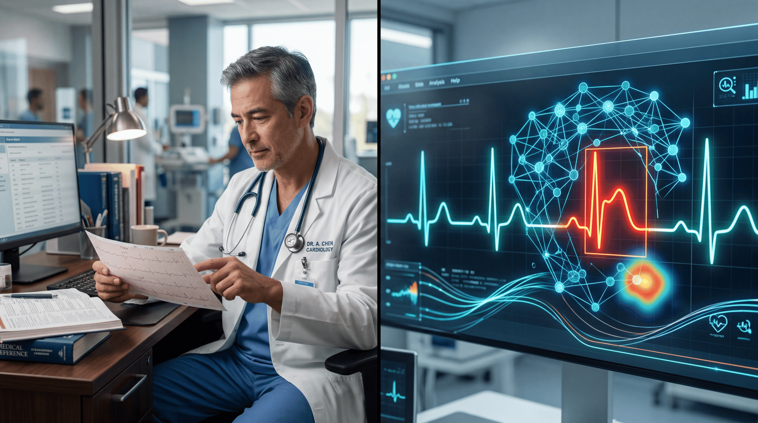

Let’s cut through the noise: AI isn’t going to replace your cardiologist, but it’s already saving lives in ways most patients don’t know about. After three decades in medicine and watching countless technology “revolutions” come and go, I can tell you that artificial intelligence in cardiac care is different—not because it’s magical, but because it’s solving specific, measurable problems that kill people every day.
The media loves to hype AI as either our healthcare savior or a dystopian nightmare. The truth is far more interesting and useful.
Where AI Actually Outperforms Human Cardiologists (And Where It Fails)
Here’s what the data shows: AI models can now interpret electrocardiograms (ECGs) with 94-97% accuracy for detecting certain arrhythmias and structural heart problems—matching or exceeding the performance of experienced cardiologists in controlled studies. That’s not hype; that’s published in the Lancet and Nature Medicine.
But here’s the mechanism that matters: these systems aren’t “thinking” about your heart. They’re pattern-matching machines trained on millions of ECG strips, identifying micro-variations in electrical signals that even skilled physicians might miss on a busy Tuesday afternoon.
The Mayo Clinic’s AI-ECG platform can detect left ventricular dysfunction—essentially a weakening heart pump—from a standard 10-second ECG with remarkable accuracy. This matters because left ventricular dysfunction often has no symptoms until it’s progressed significantly. Traditional diagnosis requires an echocardiogram (ultrasound of the heart), which costs $1,000-2,500 and requires specialized equipment.
The Biology Behind Why This Works
Your heart’s electrical system is essentially a biological circuit, and subtle disruptions in timing—we’re talking milliseconds—reflect structural or functional problems. When your left ventricle weakens, the electrical depolarization pattern shifts in ways that are mathematically detectable but visually subtle.
AI models trained on hundreds of thousands of paired ECGs and echocardiograms learn these correlations. They’re detecting patterns like slightly prolonged QRS duration, T-wave inversions in specific leads, or ST-segment changes that correlate with reduced ejection fraction (the percentage of blood your heart pumps with each beat).
This isn’t magic—it’s high-dimensional pattern recognition applied to electrophysiology. The algorithm isn’t “understanding” your heart; it’s finding statistical signatures that predict structural changes.
What’s Proven Effective Right Now (Not Five Years From Now)
Atrial fibrillation detection: Apple Watch and similar wearables using photoplethysmography (light-based pulse detection) can identify irregular heart rhythms with sensitivity around 97% and specificity of 99.6% in validation studies. The Apple Heart Study, involving over 400,000 participants, demonstrated this capability at scale.
Why this matters clinically: Atrial fibrillation (AFib) increases stroke risk five-fold. About 30% of people with AFib don’t know they have it because it’s often asymptomatic or paroxysmal (comes and goes). Early detection means earlier anticoagulation, which reduces stroke risk by about 60%.
Cardiovascular risk prediction: AI models analyzing retinal images—yes, pictures of the back of your eye—can predict cardiovascular risk with accuracy comparable to established clinical risk scores. Google’s research published in Nature Biomedical Engineering showed these models could predict age, blood pressure, and smoking status from retinal photos, then calculate 5-year cardiovascular event risk.
The mechanism: Your retinal vasculature reflects systemic vascular health. Arterial narrowing, changes in vessel tortuosity, and hemorrhages visible in the retina correlate with coronary artery disease, hypertension, and diabetes—all major cardiac risk factors.
Echocardiogram interpretation: AI algorithms can measure ejection fraction from echocardiograms faster and more reproducibly than human interpreters. Studies in Nature showed AI achieved expert-level accuracy and reduced measurement variability by 50%.
What The Media Consistently Gets Wrong
The biggest myth: AI will replace doctors. This fundamentally misunderstands how clinical medicine works. Cardiac care isn’t just pattern recognition—it’s integrating complex, often contradictory information, understanding patient preferences and social context, managing medication interactions, and making judgment calls when the data is ambiguous.
What AI does is augment specific tasks—like flagging concerning ECG patterns in emergency departments where physicians might be seeing 40 patients in a shift. It’s a sophisticated screening tool, not a replacement for clinical judgment.
Second myth: AI is always more accurate. Not true. AI models trained on predominantly white, middle-aged populations perform significantly worse on underrepresented demographic groups. A 2021 study showed that pulse oximeters—which use optical sensors similar to wearable heart monitors—overestimate oxygen saturation in Black patients by 1-2%, leading to missed diagnoses of hypoxemia.
The same bias problem affects AI cardiac models. If your training data comes primarily from academic medical centers serving relatively affluent populations, your algorithm won’t generalize to everyone. This isn’t hypothetical—it’s measured, documented, and a real problem.
Third myth: AI catches everything. AI models excel at what they’re trained for but fail catastrophically at edge cases. An AI trained to detect heart attacks from ECGs might miss pericarditis, pulmonary embolism, or hyperkalemia that presents with similar ECG changes but requires completely different treatment.
What This Means For Your Actual Heart Health
If you’re over 40 with cardiac risk factors (hypertension, diabetes, family history, smoking), AI-enabled tools make screening more accessible and affordable. That’s genuinely good news. A $300 wearable that detects AFib could prevent a stroke that would cost $50,000 to treat and leave you permanently disabled.
But here’s the critical nuance: these tools generate alerts, not diagnoses. A positive AFib screen on your smartwatch needs confirmation with a proper ECG and clinical correlation. The danger isn’t false positives per se—it’s that false positives create anxiety, drive unnecessary testing, and sometimes lead to overtreatment.
Approximately 10-15% of wearable AFib alerts are false positives. That sounds acceptable until you consider that millions of people now wear these devices. We’re generating massive numbers of worried-well patients seeking emergency evaluation for what turns out to be artifact or sinus arrhythmia.
The Future That’s Actually Coming (Based on Current Clinical Trials)
Current NIH-registered trials are testing AI for predicting sudden cardiac death, optimizing heart failure medication dosing, and identifying subclinical atherosclerosis from routine chest X-rays. These aren’t science fiction—they’re in active clinical investigation.
The most promising near-term application: AI-guided treatment optimization for heart failure. Heart failure management involves balancing multiple medications (ACE inhibitors, beta blockers, diuretics, mineralocorticoid receptor antagonists) with narrow therapeutic windows and significant drug-drug interactions. AI systems that continuously monitor symptoms, vital signs, and lab values could suggest personalized dose adjustments, potentially reducing hospitalizations by 20-30%.
What won’t happen soon: AI conducting independent diagnosis and treatment. The liability issues alone make this implausible, but more fundamentally, medicine involves explaining risks and benefits to patients, incorporating their values into treatment decisions, and handling the messy reality that most patients have multiple conditions affecting each other.
The Mechanism Problem Everyone Ignores
Here’s what keeps me up at night as a physician: most AI cardiac models are “black boxes.” They work, but we don’t fully understand why they work. When a cardiologist reads an ECG and identifies ST-segment elevation suggesting acute myocardial infarction, they can explain the pathophysiology—blocked coronary artery, ischemic myocardium, injured tissue causing electrical changes.
When an AI flags the same ECG, it’s often identifying correlations that don’t map to our understanding of cardiac physiology. Sometimes these correlations are real signals we didn’t know existed. Sometimes they’re spurious patterns in the training data. We’re often flying blind.
This matters clinically because medicine isn’t just about correct diagnoses—it’s about understanding disease mechanisms well enough to predict complications, choose between treatment options, and know when your diagnosis is probably wrong despite positive tests.
What The Guidelines Actually Say
The American College of Cardiology has issued guidance on AI in cardiovascular medicine. Key points: AI tools should augment, not replace clinical judgment. Physicians using AI tools remain responsible for clinical decisions. Patients should be informed when AI is used in their care.
The WHO’s ethics and governance of AI for health emphasizes that AI systems must be validated across diverse populations, continuously monitored for performance drift, and designed to enhance rather than replace human capabilities.
Notably, the FDA has approved over 50 AI-enabled cardiac devices, but most are designated as “decision support” tools requiring physician oversight, not autonomous diagnostic systems.
What You Should Actually Do
If you’re at elevated cardiac risk: A wearable device with FDA-cleared ECG capability (Apple Watch, AliveCor KardiaMobile) provides reasonable screening for AFib. Take any alerts seriously but don’t panic—get confirmation from your physician.
If you’re getting a routine ECG: Ask whether AI interpretation is being used and request that a physician reviews the findings. Studies show that AI-augmented interpretation (physician plus AI) outperforms either alone.
If you’re told you need extensive cardiac testing based on an AI algorithm: Ask what specific finding triggered the recommendation. “The computer said so” isn’t sufficient justification for invasive testing. Insist on understanding the clinical reasoning.
For everyone: Understand that AI in cardiac care is a tool, not an oracle. It works best for detecting specific, well-defined patterns in clean data. It works poorly with ambiguous findings, unusual presentations, or when you have multiple overlapping conditions. Your physician’s clinical judgment integrating AI findings with your complete medical picture remains essential.
The One Thing That Matters Most
AI in cardiac care represents genuine progress on specific problems—earlier AFib detection, more consistent echocardiogram interpretation, better risk stratification. These advances will save lives. But the fundamental requirements for cardiovascular health haven’t changed: control your blood pressure, manage your cholesterol, don’t smoke, maintain healthy weight, exercise regularly, control diabetes if present.
AI doesn’t replace those basics. The smartest algorithm in the world can’t save you from a lifetime of poor lifestyle choices, and the most sophisticated wearable won’t matter if you ignore the alerts or can’t access proper follow-up care. Technology is a tool, not a substitute for taking responsibility for your health and having a physician who knows you and your complete medical context.